top of page

Urgent Eyecare / Emergency 

Our experienced therapeutic optometrist has the expertise to diagnose and manage eye emergency conditions accurately and efficiently, such as foreign bodies, eye pain,  red eyes, headaches, retinal and macular emergency. Our practice is very well equipped to handle eye emergency conditions. For example, high resolution magnified photography and scans of both external and internal eye health is available to show and explain to patients for fully understanding of their conditions to improve their compliance of treatments for effective management. For those eye emergency conditions required to be managed by ophthalmologists or other medical professionals, emergency referrals and appointments can be arranged swiftly. 


Our optometrist worked in an eye specialist clinic in a hospital. He is a certified and registered as therapeutic endorsed optometrists in Australia, therefore, he is able to prescribe many eye medications to treat common eye conditions.

Some of the emergency cases have been diagnosed and managed are as below:

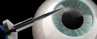
Removal of Embedded Corneal Foreign Body
A corneal foreign body is one of the main causes of blindness. The effects can range from mild infections (corneal abrasion, keratitis) to severe damage (endophthalmitis) depending on the type of material. Late-stage complications can cause permanent blindness. (Reference: The Influence of Corneal Foreign Body on Eye Infection | Atlantis Press (atlantis-press.com)

Corneal foreign body removal.

Patient: 74 year old man came in with foreign body sensation with blinkings in his right eye. Normal vision was reported.

History: A metal foreign body was found embedded in his nasal peripheral cornea outside the pupillary zone. 

Examination: High resolution magnified images were taken and used for explaining the conditions and procedures required to the prepare the patient psychologically in order to avoid panic attack; and documenting the conditions and severity for future reference and comparision.

Management: After his consent of proceeding the procedure, local anesthetic eye drops were applied and the foreign body was gently and painlessly removed with a sterilized tool. Rust residues and stain were found underneath and then successfully removed by another sterilized special tool without any discomfort.

Eye medication and instructions were given, and a review in 24 hours was scheduled to ensure normal wound healing without any complications. 

                                                                                 #3415

Guide: Local experienced therapeutic optometrists with sufficient equipment and tools, such as slit lamp imaging and foreign body removal tools, including corneal rust ring remover Algerbrush, are the best practitioners to manage these cases. All cases should return in 24 hours (or sooner if symptoms worsen) for re-evaluation to ensure the corneal wound healing normally. Untreated corneal infection and ulcers can lead to severe permanent vision loss and even loss of an eye. Close follow-up until the corneal epithelial defect resolves.

Emergency Case with Minimal Blurry Vision Of One Eye:
Papilledema Caused By A Mandarin Size Brain Tumour

Papilledema

Patient: Caucasian male, mid 20's, healthy looking.


Symptoms: Came in for a routine eye examination due to his vision in one eye was slightly blurry.

History: Personal and family medical history was normal.

Signs: Vision of the eye with the blurriness was only about 1 line less from the norm. The retinal photography showed his optic nerve heads were slightly swollen with fuzzy margin.


Management: He was advised to go to an emergency department immediately. He declined and left initially due to minimal symptoms. I called him an hour later and advised him to go an emergency department again. He listened to his friend and finally came in to get the referral and went to an emergency department. He had a scan of his brain and immediately diagnosed with a mandarin size brain tumor in his frontal lobe. He had a brain surgery 2 days later and then another brain surgery 2 days after.


Outcome: He survived the surgeries and fully recovered after a few months.

Guide: Any changes of vision, even minimal, can be caused by eye and/or visual pathway disorders, such as dry eyes, cataracts, macular, retinal, optic nerve and brain disorders, such as brain tumors.

Comprehensive eye examination is required to check eye health and visual pathway thoroughly even with slightly blurry vision. Regular eye examination is crucial because changes of vision might not be noticeable, especially with non-dominant eye and/or without fully corrected optical appliances, such as spectacles and contact lenses.

Urgent/Emergency Case With Only Occasional Mild Blurry Vision In A Routine Eye Examination     

Patient:  Female, early 50's  (AH3958)


Symptoms: TV blurry sometimes.

History: Refractive surgery for myopia correction of both eyes over 10-15 years ago. 

Signs: Reduced spectacle corrected central vision in one eye. Advanced high resolution retinal imaging and optical coherence tomography imaging with thorough analysis revealed a epiretinal memebrane and macular schisis (split).


Management: Urgent/emergency referral to retinal eye specialist. Might need eye surgery to remove the epiretinal membrane and repair the macula to prevent further damage and visual loss.

Guide: Do not ignore minor visual blur or symptoms. Seek for well equipped experienced optometrists with comprehensive eye examination and advanced testings such as high resolution retinal imaging and optical coherence  tomography imaging with thorough analysis. Many eye diseases were not be able to identified and recognised until recent advances in these types of retinal imagings because they are not usually visible on traditional standard examination. Our experienced therapeutic optometrist provides thorough examination with highest quality equipment to ensure accurate diagnosis and timely management for the best outcome for our patients.

There are cases that don't have any symptoms at all but routine eye examinations can find out one eye has severe visual impairment. Therefore, routine eye examinations and check-ups are very important. Do not ignore any advice and recommendations from professional optometrists because they are based on years of research, latest evidence and national and international professional guidelines.

Emergency Case with Unrelated Symptoms:
Advanced Proliferative Diabetic Retinopathy causing Secondary Retinal Detachment in One Eye
Advanced Cataract with Potential Sight-threatening Complications from Diabetic Retinopathy in the Other Eye
High Risk of Acute Angle-closure Glaucoma (AH4025)  

Patient:  Female, early 70's (AH4025)


Symptoms: Left eye very watery with blurry vision.

Harder to read.

Signs: Secondary retinal detachment from traction of preretinal fibrovascular tissue of advanced proliferative diabetic retinopathy in one eye. with Advanced cataract and potential sight-threatening complications caused by diabetic retinopathy in the other eye. Narrow anterior angles in both eyes; moderate high eye pressure in one eye.


Management: Refer to emergency department due to retinal detachment in the right eye, potential serious complications in the left eye and high risk of acute angle-closure glaucoma of both eyes.

Outcome: tba.

Guide: Many sight-threatening eye diseases either do not have symptoms or have very mild symptoms. It is quite often that patients come in either without any symptoms or with unrelated symptoms like this patient. Without performing a highly comprehensive eye examination, many sight-threatening or even life-threatening eye diseases can be missed. Patients often misunderstand optometrists and even ophthalmologists of over-servicing. They either decline essential in-depth testing or complain about high fees after the testing was performed. Every test recommended is essential and based on government and professional guidelines, indications and experience of practitioners.

Emergency Case With Minimal Symptoms of One Eye:
Acute Glaucoma With Very High Intra-ocular Pressure                                                

Corneal Scarring Caused By Over-the-counter Wart Remover Applied On Eyelid

Patient: Male, late 40's, healthy looking.    (AH1570)


Symptoms: Cloudy vision of left eye associated with headaches for 1 day. This happened before which was about 2 years ago; his GP had prescribed eye drops for itchiness. This time, he didn't consult GP and went to pharmacy for eye drops. The phamacist referred him to us due to his symptoms.

History: Personal and family medical history was normal.

Signs: Intra-ocular pressure: right eye 23mmHg (slightly higher than the normal limit of 21mmHg), left eye 60mmHg (extremely high!!) and this was an emergency. His whole left cornea was slightly cloudy and with global fluorescein punctate stainings. Estimated cup-disc ratio of optic nerve head: right eye 0.75, left eye 0.90.


Management: He was advised to go to an emergency department immediately due to this sight-threatening condition. He was hesitated to go due to minimal symptoms. I called him an hour later and stressed the importance of going to emergency department as soon as possible due to the possibility of permanent blindness of the left eye within a matter of days. Urgent treatments and management were crucial.

Outcome: I did not know whether he took my advice.

Guide: Do not ignore any mild visual symptoms. These can be caused by sight- and life-threatening emergency conditions which require urgent management and treatments to prevent blindness and death. Comprehensive eye examination is crucial to check eye health and visual pathway thoroughly even with minimal visual symptoms.

1420_20210511_174231_2UP_N_1342.jpg

Patient: Asian female, late 30's

Symptoms: Constant cloudy vision of left eye for 4 days.


History: This symptom started after using an over-the-counter wart remover with salicylic acid to remove a wart on her eyelid 5 days ago. The chemical went into her eye and she irrigated the eye with tap water for about 15 minutes. Then she consulted a GP and had used an antibiotic eye medication. 

Signs: The images showed a central hazy corneal scar covering most of the pupillary zone. This scar could be permanent and cause irreversible reduced vision even with spectacles or contact lenses.

Management: Corneal scar is permanent. She was advised to use non-preservative artificial tear four times a day and if needed.

Guide: Local experienced therapeutic optometrists are the best practitioners to deal with eye emergency like this, especially when the vision is affected. Because therapeutic optometrists can prescribed eye medications to treat and manage this condition, and monitor both vision and eye health. However, emergency departments are the best places for managing severe cases. 

Emergency Case With Severe Central Corneal Abrasion

Patient:  Female, early 40's (AH3824)


Symptoms: Left eye with severe pain with blurry vision after removing her disposable soft contact lens.

Signs: Severe central corneal abrasion involving pupillary zone.


Management: Lay-off contact lens. Prescription (non over-the-counter) antibiotic eye drops for contact lens wearer were prescribed. Refer to see ophthalmologist urgently. A few visits were required to monitor the progress.

Outcome: Cornea healed gradually over weeks and vision was still affected. Spectacle prescription changed and new lenses were required.

Guide: Many contact lens wearers, especially disposable lenses, have poor compliance or even ignore hygiene, source of lenses and solution (if any), regular lens replacements and checkup schedules. In fact, they are exposing themselves to the risks of permanent severe visual impairement. Contact lens wear is very safe if professional instructions and ongoing

guidance  and are followed. However, updated 

information and guidance cannot be obtained from the practitioners if checkups are ignored or avoided. In fact, any eye problems arised might 

cause pain, inconvenience, substantial cost to manage and treat them. In some cases, visual impairment can affect their independence, life style, job, income and future. Simply following professional advice is the best way to avoid all these issues.

Emergency Case Without Related Symptoms:
High Intracranial Pressure (AH1322) - Information coming soon.                                          

Emergency Case With Multiple Symptoms:
High Intracranial Pressure (AH3539) - Information coming soon.                                               

bottom of page